Wrist Pain
A painful wrist can be a diagnostic and therapeutic challenge. Many pathologies affect the wrist, however a history and careful examination can often establish the diagnosis as signs are usually well localised.
It is useful to consider groups of pathologies based on their location. Common pathologies for each location are listed:
(1) Radial sided Pathology: De Quervain’s Tenosynovitis (proximal to the radial styloid), Osteoarthritis at the first carpometacarpal joint (CMCJ OA) (base of thumb), Scaphoid pathology (snuffbox).
(2) Dorsal / Central Pathology: Dorsal wrist ganglion, Kienbock’s disease (Lunate osteonecrosis), ligament wear and tear between Scaphoid and Lunate.
(3) Ulnar sided Pathology: Distal Radial-Ulnar joint (DRUJ) arthritis, Triangular Fibro-cartilage (TFCC) wear or tear, Extensor Carpi Ulnaris tendonitis.
Tips:
History:
- It is important to establish any history of trauma, however trivial or historical.
- Insidious onset base of thumb pain is most likely to be thumb CMCJ OA, with arthritis of the thumb being as frequent as that of the hip.
- Wrist pain after a new baby is almost pathognomonic of De Quervain’s.
Exam:
- Ask the patient to point (with one finger only) to the exact point of maximal tenderness.
- Gently palpate the wrist clockwise or counter-clockwise starting opposite to the site of pain.
- Tendonitis is commonest on the radial side (De Quervain’s) however any of the 23 tendons crossing the wrist can become inflamed.
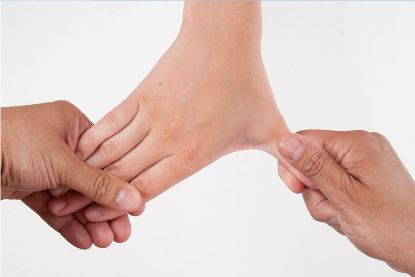
- Finklestein’s test is excellent for De Quervain’s but is often confused with Eichoff’s test (which is not as specific). It is correctly performed by grasping the patients thumb and sharply ulnarly deviating the hand, inducing pain on the radial side of the wrist (Fig.1).
First Line Investigations:
- Plain X-ray can be very useful. Look for scaphoid pathology and base of thumb arthritis on the radial side; lunate pathology as well as established wear between scaphoid and lunate may show up centrally; DRUJ arthritis and TFCC wear can often be identified on the ulnar side.
- Ultrasound is effective in identifying wrist ganglions and establishing suspected tendonitis.
Early management
- Simple analgesia, NSAIDS and splintage are appropriate modalities to manage wrist pain early on.
- A well fitted Futura type Velcro splint is ideal for cosmesis and hygiene
- Encourage patients to mobilise the rest of the upper limb including neck, shoulder, elbow and particularly fingers to avoid secondary stiffness.
- Need for on going splintage following a minor injury warrants further investigations (as described above).
When to refer?
Chronic wrist pain: Continued pain in the absence of a diagnosis, despite the above investigations and management, warrants further, second line investigation by way of MRI and onward referral to a hand and wrist specialist. Wrist arthroscopy is indicated in unexplained chronic pain for over three months.
What can be done? Wrist arthroscopy technology has advanced considerably. This allows us to visualise the entire wrist joint and is the diagnostic gold standard. Modern instrumentation and techniques allow us to access, repair and debride areas of pathology particularly on the ulnar side of the wrist with reliable outcomes. TFCC pathology, loose bodies, synovectomy and excision of dorsal wrist ganglion can be addressed.

Fig.1: The correct Finklestein’s test is far more specific for De Quervain’s.


